Beranda
/ Mesothelioma Histology Tonofilaments / Malignant And Borderline Mesothelial Tumors Of The Pleura Sciencedirect / Histopathology is the study of diseased cells.
Mesothelioma Histology Tonofilaments / Malignant And Borderline Mesothelial Tumors Of The Pleura Sciencedirect / Histopathology is the study of diseased cells.
Insurance Gas/Electricity Loans Mortgage Attorney Lawyer Donate Conference Call Degree Credit Treatment Software Classes Recovery Trading Rehab Hosting Transfer Cord Blood Claim compensation mesothelioma mesothelioma attorney Houston car accident lawyer moreno valley can you sue a doctor for wrong diagnosis doctorate in security top online doctoral programs in business educational leadership doctoral programs online car accident doctor atlanta car accident doctor atlanta accident attorney rancho Cucamonga truck accident attorney san Antonio ONLINE BUSINESS DEGREE PROGRAMS ACCREDITED online accredited psychology degree masters degree in human resources online public administration masters degree online bitcoin merchant account bitcoin merchant services compare car insurance auto insurance troy mi seo explanation digital marketing degree floridaseo company fitness showrooms stamfordct how to work more efficiently seowordpress tips meaning of seo what is an seo what does an seo do what seo stands for best seotips google seo advice seo steps, The secure cloud-based platform for smart service delivery. Safelink is used by legal, professional and financial services to protect sensitive information, accelerate business processes and increase productivity. Use Safelink to collaborate securely with clients, colleagues and external parties. Safelink has a menu of workspace types with advanced features for dispute resolution, running deals and customised client portal creation. All data is encrypted (at rest and in transit and you retain your own encryption keys. Our titan security framework ensures your data is secure and you even have the option to choose your own data location from Channel Islands, London (UK), Dublin (EU), Australia.
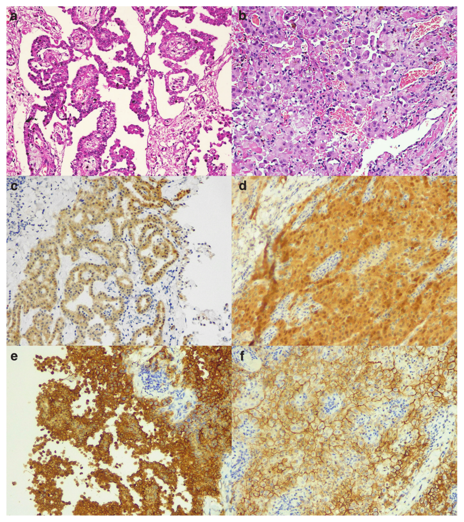
Mesothelioma Histology Tonofilaments / Malignant And Borderline Mesothelial Tumors Of The Pleura Sciencedirect / Histopathology is the study of diseased cells.. Consequently, this case was diagnosed as deciduoid mesothelioma and 2 years and 4 months after operation, the patient's clinical course has been good. Fixation for em is often done with glutaraldehyde. In the present study, however, ck 5/6 was not found useful in the diagnosis of mesothelioma since it labelled. Mesotheliomas could be distinguished from adenocarcinomas of the lung by length of microvilli (mean ratios of length to diameter ldr, 15.7 and 8.7. The adenomatoid tumor is uniformly benign, whereas the diffuse malignant mesothelioma pursues a downhill clinical course, rapidly leading to fatality.
It is commonly abbreviated em. In a 2019 case study, the patient claimed to have lung cancer, but proper pathology scans showed mesothelioma cancer cells, not lung cancer cells. 2.(a) the surface of omentum showed multiple white, firm nodules. Ultrastructurally, numerous, long microvilli, tonofilaments and desmosome junctions could be seen. Consequently, this case was diagnosed as deciduoid mesothelioma and 2 years and 4 months after operation, the patient's clinical course has been good.
Cancers Free Full Text Primary Ovarian Mesothelioma A Case Series With Electron Microscopy Examination And Review Of The Literature Html from www.mdpi.com A moderate number of slender microvilli was also present (fig. Histology is a branch of biology that involves the study of cells and tissues. Bap1 is lost in the majority of epithelioid mesotheliomas and is indicative of malignancy but is not a sensitive initial diagnostic marker. Ultrastructurally, the neoplastic cells had prominent surface microvilli, numerous desmosomes, and tonofilaments. Searching for mesothelioma tonofilaments searching for mesothelioma tonofilaments benign cystic mesothelioma histology hipec for peritoneal mesothelioma is mesothelioma a form of cancer fak inhibitor mesothelioma exotic carts mesothelioma outbreak life expectancy rate mesothelioma hodgkin lumphoms mesothelioma hx family mesothelioma how long does it take to die from mesothelioma how to get. It is commonly abbreviated em. A, mesothelioma with ,numerous long, thin microvilli projecting frot'n the surface [ratio ofilength to diameter, 16.4). Immunohistochemical and ultrastructural findings supported the mesothelial nature of the tumor, and led us to diagnose this tumor as a sarcomatoid localized malignant mesothelioma.
Mesothelioma associated with and without asbestosis is considered rare.
This case is considered to be the first reported in japan. Physicians often fail to say the difference between pleural mesothelioma and lung cancer. A, mesothelioma with ,numerous long, thin microvilli projecting frot'n the surface [ratio ofilength to diameter, 16.4). The adenomatoid tumor is uniformly benign, whereas the diffuse malignant mesothelioma pursues a downhill clinical course, rapidly leading to fatality. Histology is a branch of biology that involves the study of cells and tissues. Pubmed pubmedcentral crossref google scholar Consequently, this case was diagnosed as deciduoid mesothelioma and 2 years and 4 months after operation, the patient's clinical course has been good. Histology is a branch of biology that involves the study of cells and tissues. 2.(a) the surface of omentum showed multiple white, firm nodules. Tonofilaments were identified by electron microscopy in one of the cases. Histopathologically, a biopsy specimen from the retroperitoneal tumor revealed a biphasic type of malignant mesothelioma. A history of asbestos exposure was obtained from three patients. A similar hypothesis was proposed by carey et al on identifying both focal tonofilaments, usually found only in mesothelial cells, and abortive cilia, indicating müllerian differentiation, in multiple focal ovarian serous carcinoma.
Primary serosal neoplasms demonstrate a wide spectrum of growth patterns and biologic aggressiveness. Mesothelioma tissues have the singular potential of producing tumors of epithelial or mesenchymal type, or both. Physicians often fail to say the difference between pleural mesothelioma and lung cancer. Histology (biphasic and sarcomatoid) lymph node metastasis. Other findings indicating mesothelial lineage of the malignant cells are filaments, which often surround the nucleus, and cytoplasmic, coarse tonofilaments, the latter correlating to the expression of cytokeratin 5 and a finding of squamoid differentiation.
2 from Six examples of malignant mesothelioma appearing as a localized pleural mass are described. Tonofilaments were much more figure i. A similar hypothesis was proposed by carey et al on identifying both focal tonofilaments, usually found only in mesothelial cells, and abortive cilia, indicating müllerian differentiation, in multiple focal ovarian serous carcinoma. Fixation for em is often done with glutaraldehyde. Malignant mesothelioma in situ could be an additional category routinely grade epithelioid malignant pleural mesothelioma. Histopathology is the study of diseased cells. Carlton experimental pathology department, medical research division, american cyanamid company, pearl river, new york, and department of veterinary pathobiology, college of veterinmy medicine, purdue university, west lafayette, indiana, u.s.a. Electron microscopy was also performed.
Combined quantitative and qualitative features were evaluated to provide criteria for distinguishing among these three tumors, which may present as either primary or metastatic pleural tumors.
Mesotheliomas could be distinguished from adenocarcinomas of the lung by length of microvilli (mean ratios of length to diameter ldr, 15.7 and 8.7. A history of asbestos exposure was obtained from three patients. In the present study, however, ck 5/6 was not found useful in the diagnosis of mesothelioma since it labelled. Ultrastructurally, numerous, long microvilli, tonofilaments and desmosome junctions could be seen. Six examples of malignant mesothelioma appearing as a localized pleural mass are described. The tumors varied histologically, but the most common type was epithelial with a papillary pattern. Tonofilaments showing many short fascicles with various 109,43337 naturally occurring atriocaval mesotheliomas in rats m. The cystic peritoneal mesothelioma occupies an … Six examples of malignant mesothelioma appearing as a localized pleural mass are described. The tumors ranged in size from 2.8 to 10 cm. There were four women and two men, ranging in age from 42 to 76 years. Histology is a branch of biology that involves the study of cells and tissues.
Electron microscopy disclosed that the tumor cell … Physicians often fail to say the difference between pleural mesothelioma and lung cancer. Histopathology falls within the larger field of pathology. Pubmed pubmedcentral crossref google scholar A similar hypothesis was proposed by carey et al on identifying both focal tonofilaments, usually found only in mesothelial cells, and abortive cilia, indicating müllerian differentiation, in multiple focal ovarian serous carcinoma.
Reactive Mesothelial Hyperplasia Mimicking Mesothelioma In An African Green Monkey Chlorocebus Aethiops Abstract Europe Pmc from europepmc.org Of tonofilaments were seen within the cytoplasm. A case of peritoneal malignant mesothelioma in a radiation technologist, who had worked in this field for 34 years, is reported. In human pathology, ck 5/6 has high sensitivity and specificity for epithelial mesothelioma and is now recognized as a valuable marker in distinguishing this neoplasm from pulmonary adenocarcinoma (attanoos et al., 2002; Histology is a branch of biology that involves the study of cells and tissues. Tonofilaments frequently in a perinuclear distribution molecular / cytogenetics description. Ultrastructurally, numerous, long microvilli, tonofilaments and desmosome junctions could be seen. There were four women and two men, ranging in age from 42 to 76 years. Combined quantitative and qualitative features were evaluated to provide criteria for distinguishing among these three tumors, which may present as either primary or metastatic pleural tumors.
Tonofilaments were much more figure i.
2.(a) the surface of omentum showed multiple white, firm nodules. Ultrastructurally, numerous, long microvilli, tonofilaments and desmosome junctions could be seen. The cystic peritoneal mesothelioma occupies an … Flow cytometry showed an aneuploid dna content in four tumors and a diploid content in one. Mesothelioma associated with and without asbestosis is considered rare. Carlton experimental pathology department, medical research division, american cyanamid company, pearl river, new york, and department of veterinary pathobiology, college of veterinmy medicine, purdue university, west lafayette, indiana, u.s.a. The adenomatoid tumor is uniformly benign, whereas the diffuse malignant mesothelioma pursues a downhill clinical course, rapidly leading to fatality. Histology is a branch of biology that involves the study of cells and tissues. Mesothelioma with asbestosis is so rare that it is worth. Gata3 and p16 are not specific to mesothelioma. A history of asbestos exposure was obtained from three patients. Cell type of malignant mesothelioma histology Mesothelioma tissues have the singular potential of producing tumors of epithelial or mesenchymal type, or both.
Introductiona recurrent problem in diagnostic pathology is the inability to reliably distinguish pleural malignant mesothelioma (mm) from pleural involvement by metastatic carcinoma, particularly pulmonary adenocarcinoma (ac) 1, 2this may be a very difficult task on the basis of routine histology alone and the problem is often compounded by incomplete clinical evaluation and limited mesothelioma histology. Ultrastructurally, the neoplastic cells had prominent surface microvilli, numerous desmosomes, and tonofilaments.